Scanning electron microscope images of beaded Sepharose CL-6B with 100
Par un écrivain mystérieux
Last updated 15 juillet 2024


Sepharose CL-6B Cross-linked 62610-50-8

Scanning electron microscope images of beaded Sepharose CL-6B with 100

IJMS, Free Full-Text
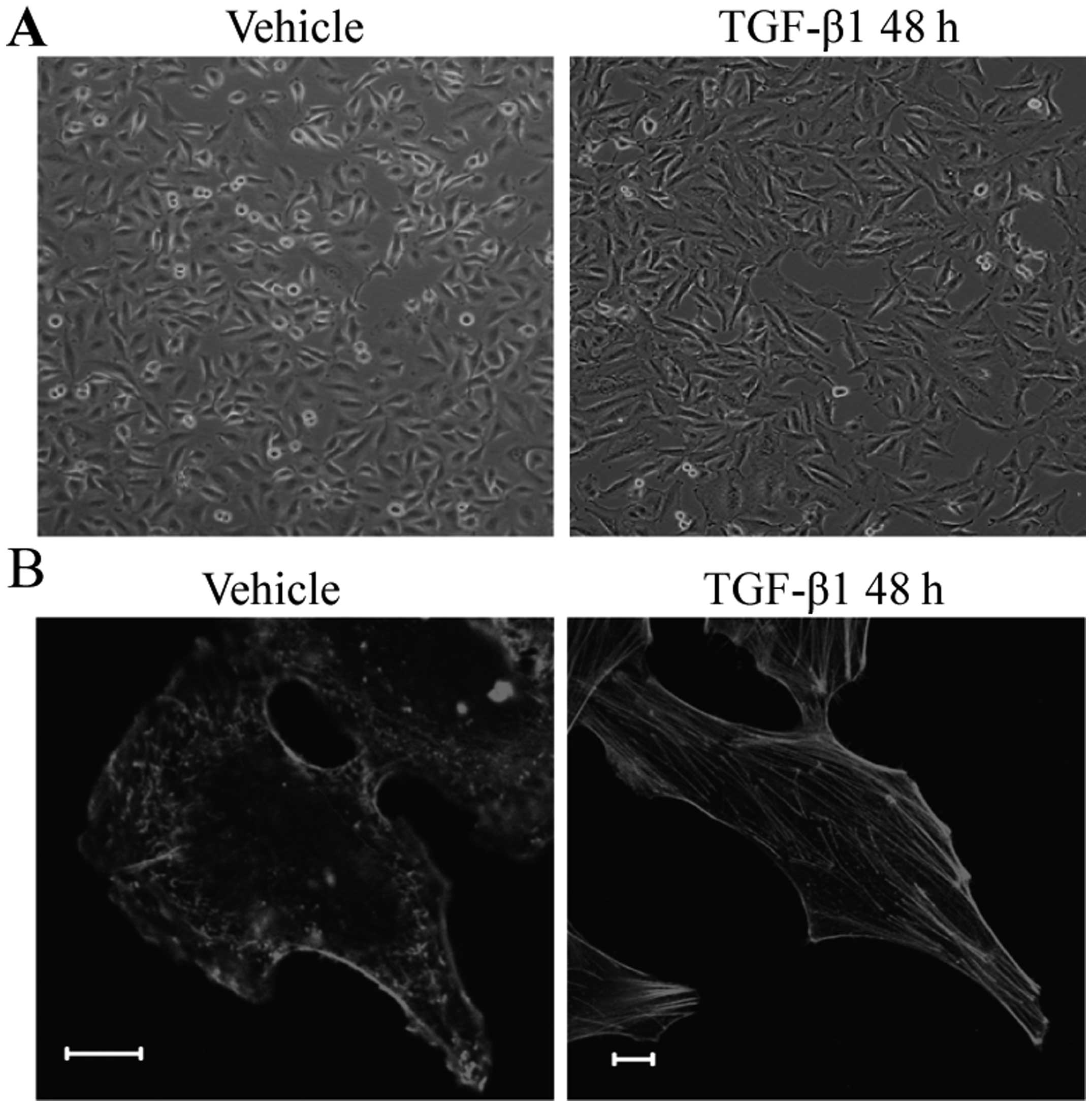
Ezrin is required for epithelial-mesenchymal transition induced by TGF-β1 in A549 cells

Impact of mechanical compression achieved by multi- ple incremental

Activation mechanism dependent surface exposure of cellular factor XIII on activated platelets and platelet microparticles - Somodi - 2022 - Journal of Thrombosis and Haemostasis - Wiley Online Library

Affinity-Based Magnetic Particles for the Purification of Single-Stranded DNA Scaffolds for Biomanufacturing DNA-Origami Nanostructures
A perspective on the isolation and characterization of extracellular vesicles from different biofluids - RSC Advances (RSC Publishing) DOI:10.1039/D1RA01576A

Scanning electron microscope images of beaded Sepharose CL-6B with 100
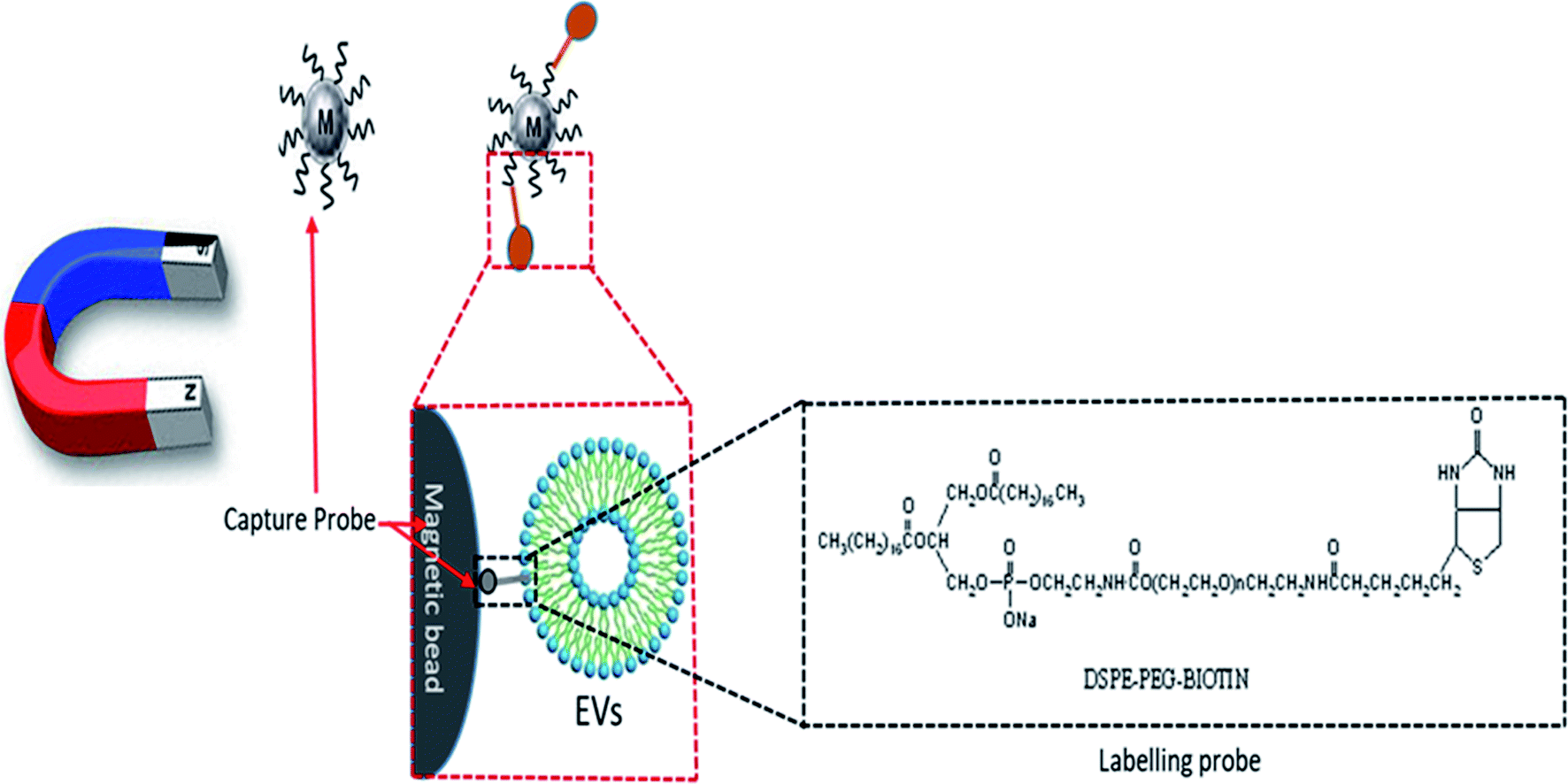
A perspective on the isolation and characterization of extracellular vesicles from different biofluids - RSC Advances (RSC Publishing) DOI:10.1039/D1RA01576A

Scanning electron microscope images of beaded Sepharose CL-6B with 100

Dual-Mode and Label-Free Detection of Exosomes from Plasma Using an Electrochemical Quartz Crystal Microbalance with Dissipation Monitoring
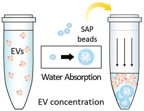
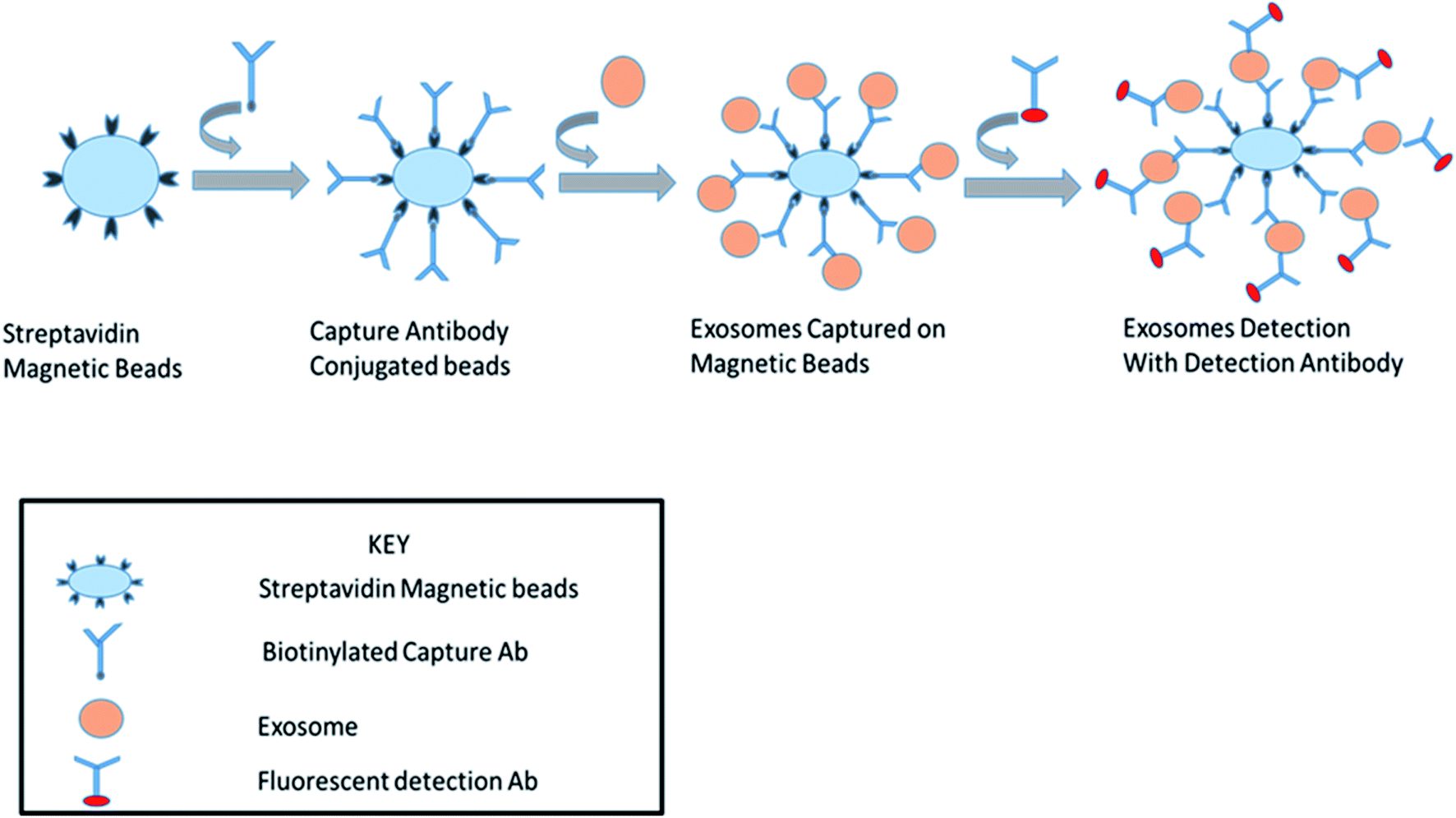
Frontiers MicroRNAs in extracellular vesicles: Sorting mechanisms, diagnostic value, isolation, and detection technology

Preparation of DAMDPA-activated Sepharose CL-6B by periodate oxidative
Recommandé pour vous
 Carson MP-250 100x ~ 250x Pocket Microscope Magnif1er MicroFlip MP250 Viewer14 Jul 2023
 Amazing Sights In Your Own Home — The Carson MP-250 LED / UV Lighted Pocket Microscope (MicroFlip™)14 Jul 2023
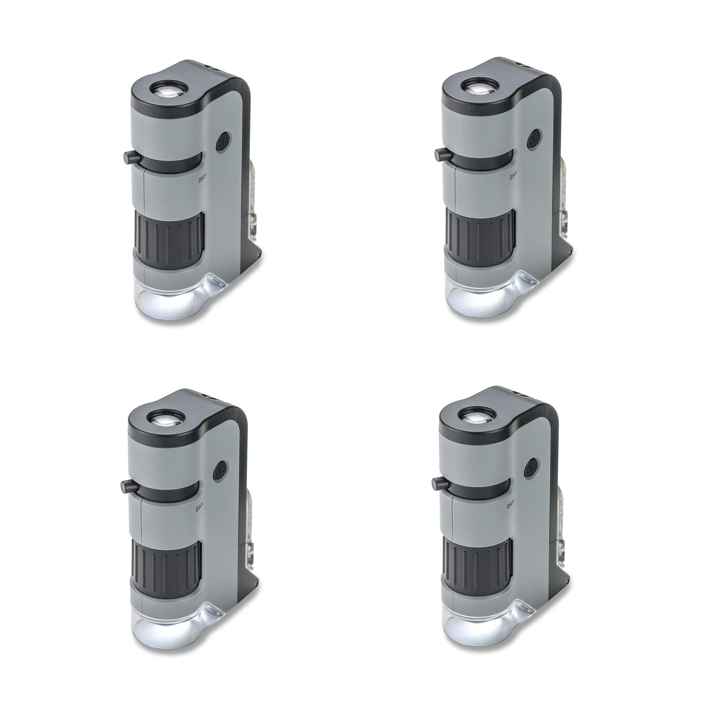 Carson 100x-250x MicroFlip LED Microscope (4 Pack)14 Jul 2023
 AmScope 40X-2500X USB-C Rechargeable Student Binocular Compound Microscope w/3D Two-Layer Mechanical Stage, Achromatic Objective - AliExpress14 Jul 2023
 Alga HD Microscope 100/250/500x, På lager14 Jul 2023
 Carson MicroFlip 100x-250x LED and UV Lighted Pocket Microscope with Flip Down Slide Base and Smartphone Digiscoping Clip (MP-250) : Industrial & Scientific14 Jul 2023
 Microscope For Adults Kids Students 100-2000x Magnification Powerful Biological Educational Microscopes14 Jul 2023
 BioBlue Series Compound Microscope, Trinocular with Camera, SMP, 4/10/S40/S100x Oil Objectives with Mechanical Stage14 Jul 2023
 291X-7280X Electron Compound Lab Microscope 12 Million Pixels USB2.0 Camera14 Jul 2023
 Herwicm Microscope for adults40-2000XCompound Microscope,Dual LED Illumination for School Home Lab Educational Gift Child Student Beginner Microscope : Electronics14 Jul 2023
Tu pourrais aussi aimer
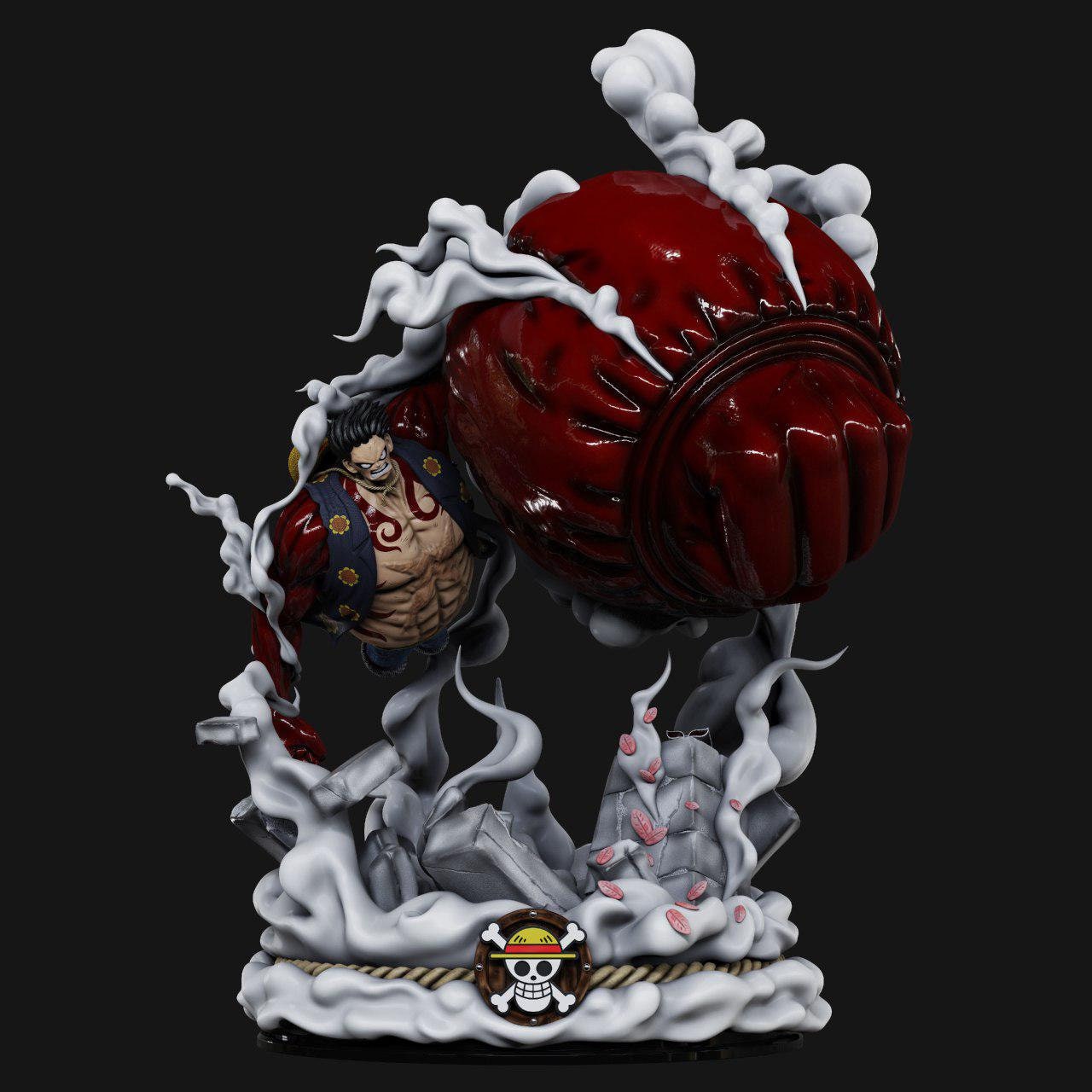 One Piece Luffy Gear 4 3D Print Model14 Jul 2023
 Plein d'idées pour réussir son déguisement année 80 Deguisement disco, Déguisement années 80, Look disco14 Jul 2023
 Gourmet Steel - Petite Poêle Inox Induction - 20cm - Revêtement14 Jul 2023
 Quelle taille choisir pour une taie de traversin ? • Blog14 Jul 2023
 Toni en famille DVD - Nathan Ambrosioni - DVD Zone 2 - Achat & prix14 Jul 2023
 Ajustement Pirate Bras Nivellement Lifter Outil Auxiliaire14 Jul 2023
 Chapeau Hat Pure Cotton Turban14 Jul 2023
 Coque téléphone 3D Stitch – UNICORNDOLL14 Jul 2023
 Finish Maroc. Finish Pastilles Lave Vaisselle Powerball Classic 90 Tablettes .Meilleur prix en ligne Maroc ,Marrakech,Casablanca14 Jul 2023
 Répulsif Ultrason Anti-Moustique, Anti-Rongeurs Insectes Répulsif14 Jul 2023