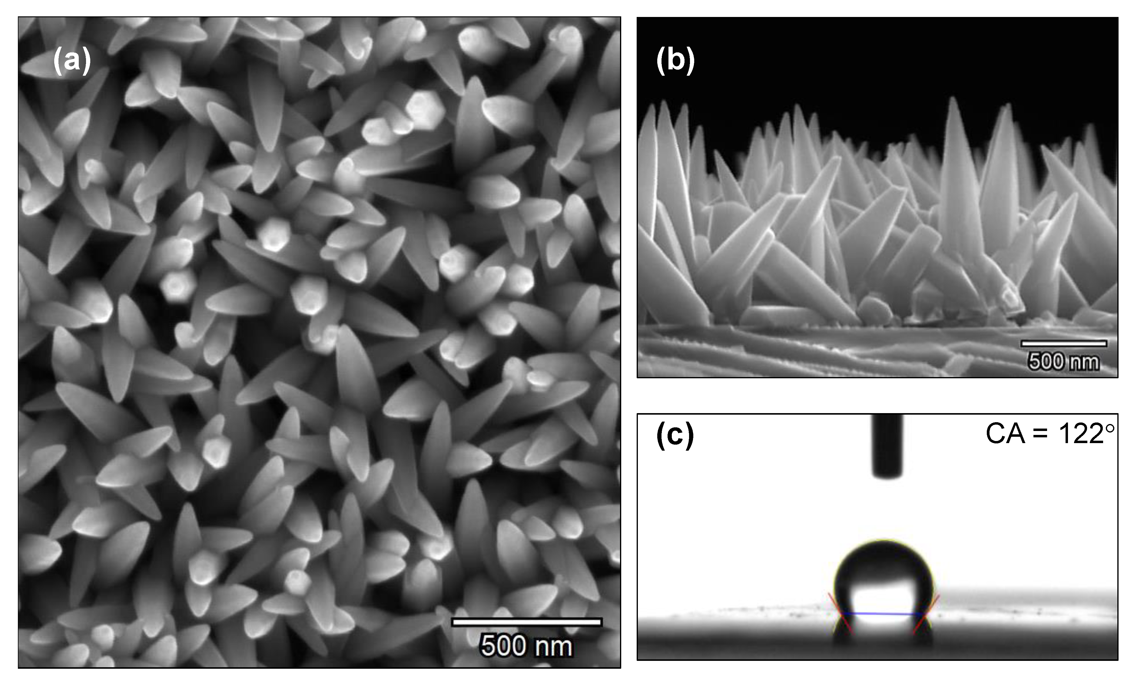
Scanning Electron Microscope Image of Zinc oxide nano rods (100-250 nm).
Par un écrivain mystérieux
Last updated 09 juillet 2024

Download scientific diagram | Scanning Electron Microscope Image of Zinc oxide nano rods (100-250 nm). from publication: Applications of Zinc Oxide Nanorods as Photocatalyst for the Decontami-nation of Imidacloprid and Spirotetramat Residues in Water | Zinc oxide nanorods having the size 100 to 250 nm and 1 to 2 m length were prepared by reacting zinc acetate with triethanolamine. The structure of the nanorods was confirmed by scanning electron microscope analysis. Photocata-lytic activity of zinc oxide nanorods on the new | Nanorods, Zinc Oxide and Zinc | ResearchGate, the professional network for scientists.

Zinc oxide nanoparticles from microwave-assisted solvothermal

Hydrothermally grown molybdenum doped ZnO nanorod arrays. The
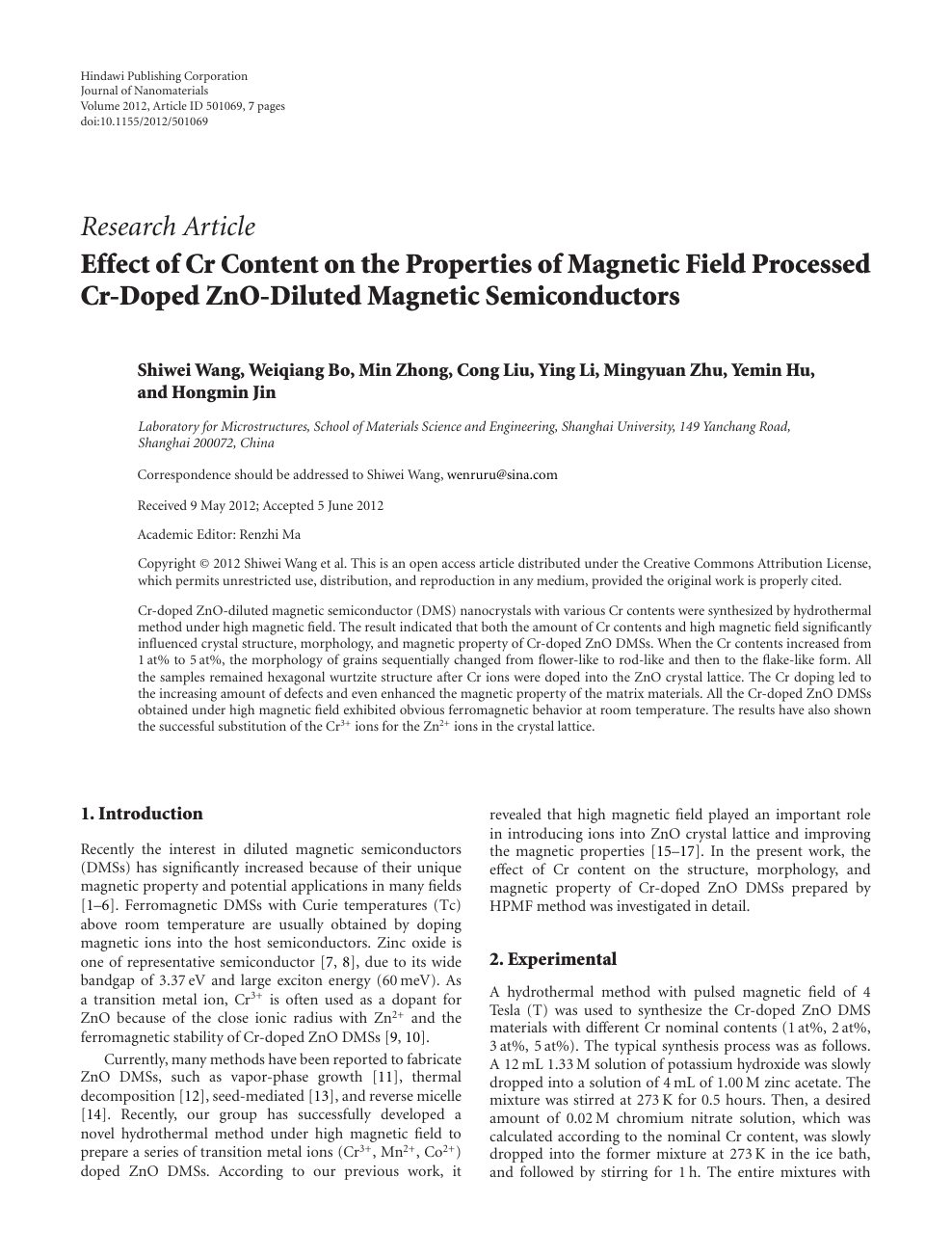
Effect of Cr Content on the Properties of Magnetic Field Processed

Electron-microscope images of the zinc oxide nanorods of different

The effect of annealing time and temperature on morphology and
Nanomaterials, Free Full-Text

Synergistic effect of coconut milk and water on synthesizing zinc
Characterization of Zinc Oxide Nanorod Samples

PDF) Structural and optical characterization and efficacy of

Frontiers Effects of Fe self-ion irradiation on a low carbon MX

Scanning electron microscope images of zinc oxide nanorods

File:Zinc oxide nanoparticles.png - Wikipedia
Recommandé pour vous
 MicroFlip™ 100x-250x LED UV Pocket Microscope with Smartphone Clip14 Jul 2023
 BioBlue Series Compound Microscope, Binocular, SMP, 4/10/S40/S100x Objectives with Mechanical Stage14 Jul 2023
 Microscope Advanced - Binocular — Eisco Labs14 Jul 2023
 Dual-Viewing Vertical Teaching Head Microscope with Mechanical Stage & 100x - LED14 Jul 2023
 Scanning electron microscope images of beaded Sepharose CL-6B with 10014 Jul 2023
 40X-2500X Super Speed USB3 18MP Digital Darkfield Trinocular LED Lab M – AmScope EU14 Jul 2023
 MAXLAPTER Microscope for Adults Kids Students 100-2000x Powerful Biological Educational Microscopes with Operation Accessories (10p), Slides Set (15p), Phone Adapter, Wire Shutter & Backpack Wr855(white)14 Jul 2023
 Premium Photo Woman doctor examines through a microscope laboratory research with a microscope14 Jul 2023
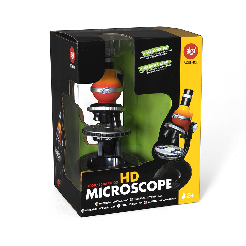 Alga Science - HD Microscope, 100/250/500X14 Jul 2023
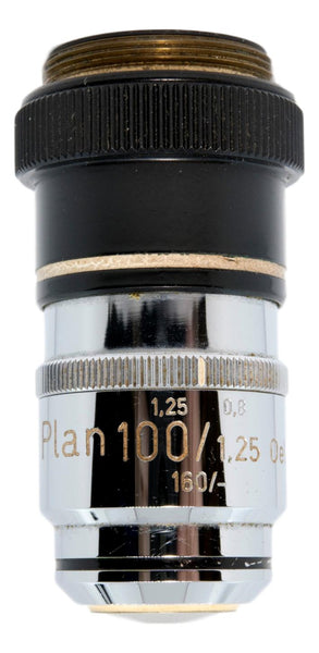 Zeiss 100x Plan Correction-Collar Oil Objective – Microscope Central14 Jul 2023
Tu pourrais aussi aimer
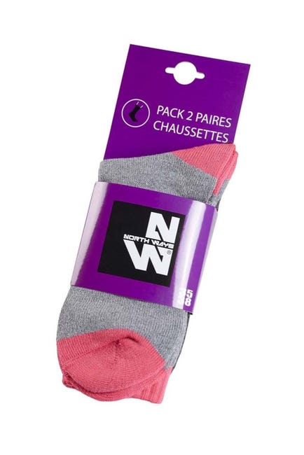 Lot de 2 paires de chaussettes de travail femme, gris/rose taille 39-4214 Jul 2023
 KIT DE MAISON de poupée bricolage fait à la main avec des meubles14 Jul 2023
 Haakaa Bebe Fruits Alimentaire Feeder & Mini Rwanda14 Jul 2023
 Einhell Aspirateur-balai sans fil TE-SV 18 Li-Solo Power X-Change (Li-Ion, 18 V, sans sac, réservoir de 0,6 L, technologie cyclonique, système triple filtration) : : Cuisine et Maison14 Jul 2023
 Tirelire électronique Coffre-fort Boîte d'économie d'argent Jouets14 Jul 2023
 Sacoche plate noire unie avec crocodile métallique et un aspect piqué LACOSTE VÊTEMENT - CCV Mode14 Jul 2023
 Matelas BEBE Primo 60x120 cm14 Jul 2023
 1 Pièce Éplucheur Multifonctionnel En Acier Inoxydable Pour Fruits Et Légumes, Ustensile De Cuisine, Mode en ligne14 Jul 2023
 Tapis de motricité sonore et lumineux Trottibul Oxybul pour enfant14 Jul 2023
 Sifflets ultrason Blackway noir - Pièces Carénage sur La Bécanerie14 Jul 2023